Eye Anatomy - Why Our Eye Are So Amazing
We learn in school that sight is one of our five senses along with, hearing, touch, taste and smell, all of which are important to help us process the world around us, but of all the five senses it is sight people fear losing the most. According to fightforsight.org, a huge 78% of people fear losing their sight the most.
Surprisingly many of us don't know much about the eye other than what we see, most people know what the eyelids and eyelashes are for and are familiar with irises, pupils and the conjunctiva (the whites of the eyes) but there is so much more that is hidden, yet important both for vision and eye health.
How Does The Eye Work?
It has been said that the eyes function very much like a camera, but to be more accurate, a camera functions very much like the human eye.
To process what we see the light is passed through the pupil via the cornea, then light passes the the lens and together they focus the light on the retina at the back of the eye. Photoreceptors then turn this light into electrical impulses that can travel from the retina via the optic nerve to the brain which interprets the signals into an image.
Anatomy Of The Eye
When we visit an Optometrist for an eye exam they look beyond the surface of the eye to determine eye health and diagnose medical conditions, so what exactly are they looking at, and what are they looking for?
By using our image of the anatomy of the eye we can help explain the various significant parts of the eye and their functions.
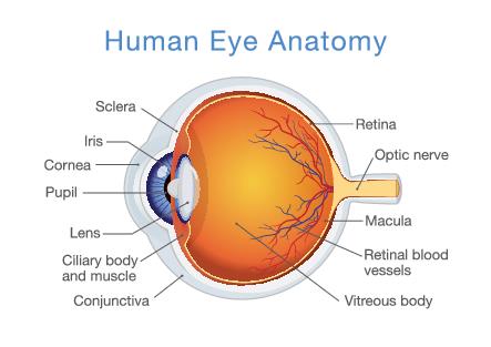
Anatomy Of The Human Eye
Sclera
The Sclera is the white part of the eye that you can see, but also surrounds the eye, covering over 80% of the eye's surface.
The sclera is a tough and fibrous tissue that provides a barrier, helping to prevent injury to the inner eye and along with Interocular pressure from the fluid within the eye, helps the eye keep its shape.
The colour of the sclera can help determine some medical conditions, a yellowing of the sclera may indicate Jaundice or hepatitis, and a bluish colour may indicate Marfan's disorder or brittle bone disease.
A 'red eye' from conjunctivitis is not actually the sclera that is red but the conjunctiva.
Cornea
The cornea is a clear layer that covers and protects the iris and the pupil allowing light to enter, it also provides between 65% and 75% of the focusing power of the eye.
The cornea is one of the most sensitive tissues in the body, consisting of 5 layers and is approximately 0.5mm thick, it is the only part of the body that does not have its own blood supply, receiving its oxygen requirements directly from the air, which is why the oxygen permeability of a contact lens is so important.
There are various conditions that can affect the cornea such as keratoconus, corneal abrasions and corneal ulcers (which we have discussed at length in one of our more recent blog posts). Dry eye syndrome can also affect the cornea causing discomfort and vision disturbances.
Iris
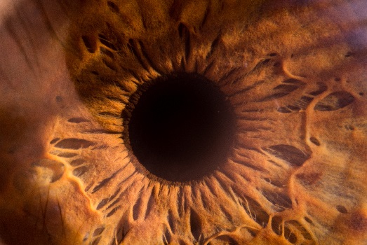
The iris is the coloured part of the eye. It is actually a disc shaped membrane with an opening in the centre that sits behind the cornea, along with the pupil the iris controls the amount of light entering the eye. It appears as though our pupil dilates and contracts but actually it is the iris doing all the work, altering the aperture of the pupil, hence controlling the amount of light entering the eye.
Rightly or wrongly the iris gets most of the attention, it is one of our most distinguished features, when we describe someone it is rare to leave out the colour of their eyes, yet rarely do we describe their shape or length of eyelashes. Your iris colour and pattern is more unique than your fingerprint and in close-ups look like a work of art.
Pupil
The pupil is the black hole at the centre of the eye and contracts or dilates depending on the amount of light required to process an object. It can only do this because of the muscular nature of the surrounding Iris that effectively opens or closes the aperture of the pupil.
It is not only light or lack of it that makes our pupils dilate, pleasure, surprise or concentration can all cause our pupils to dilate, pupil size can also be used to investigate a range of psychological phenomena. Drugs or medication can also cause the pupil to constrict or dilate. We have all seen films where a Doctor shines a light into a person's eyes - they are trying to see if there is a normal pupil reaction, as an abnormal one can indicate trauma to the brain like a stroke or aneurysm.
Lens
The lens is a smooth transparent disc shaped structure situated behind the pupil and iris, by changing its shape it can bring objects in to focus on the retina at the back of the eye. With the assistance of the cornea the lens can refract or bend the light.
The lens of the eye is approximately 10 mm across and 4 mm deep, although this is constantly altering as it adjusts its shape to bring objects into focus. The lens needs to be transparent to enable us to see clearly. A cataract causes the lens to become increasingly opaque causing eyesight to become cloudy or misty. This can also affect the colours that we see.
Monet's paintings were noticeably affected by the progression of his cataracts. The effects of the cataracts could be seen in his work, as Monet simply painted what he saw. His brush strokes became broader as his vision became blurrier. His whites, greens, and blues begin to change shade and disappear, replaced by yellows and purples.
Ciliary Body
The ciliary body is a circular structure that is an extension of the iris. It has two main functions, to produce the aqueous humor - the fluid within the eyeball, and to help change the shape of the lens via the ciliary muscle, a process known as accommodation.
Conjunctiva
The conjunctiva is a thin transparent layer of tissue covering the sclera (whites of the eye) and the inside of the upper and lower eyelids, it does not cover the cornea. Its primary function is to keep the surface of the eyes and the inner eyelids moist, it does this by producing mucus and tears, it also helps eradicate dust and small foreign objects from the eye. When the conjunctiva becomes inflamed either through infection or allergies this is called conjunctivitis or pink eye.
Retina
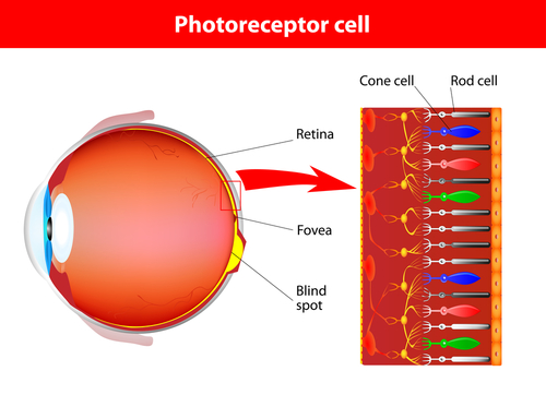
The retina is a thin multi-layered membrane covering approximately 65% of the back of the eye and plays a fundamental part in enabling us to see the world around us. With the use of photoreceptors called rods and cones, the light received via the lens is converted into chemical and nervous impulses that are relayed to the brain for interpretation via the optic nerve.
Optic Nerve
The optic nerve is situated at the back of the eye, it connects the eye to the brain. Medically referred to as the second cranial nerve or CNII, its function is to transfer information from the rods and cones in the retina using electrical impulses to the visual cortex of the brain, where the impulses are converted into an image.
Optometrists and Doctors can learn a lot about your health by looking at the optic nerve. For example, a pale optic nerve can indicate the onset of multiple sclerosis or glaucoma.
Macular
The macular is a part of the retina and situated at the back of the eye, whilst the retina concentrates on our peripheral vision this small 5mm layer of tissue is fundamental for our central vision, much of our colour vision and distinguishing fine details.
In the centre of the macular is a blood vessel-free spot called the fovea.
If the macular deteriorates then we lose our ability to see clearly in our central vision making tasks like reading, driving and identifying faces difficult.
Retinal Blood Vessels
The central retinal artery enters the eye via the optic nerve, before splitting in two forming the superior and inferior (upper and lower) vessels. These vessels then divide further and further, getting progressively smaller and smaller each time, ending in capillaries, similar to the branches of a tree. It is these arteries, vessels and capillaries that make up the retinal blood vessels that bring oxygenated blood and nutrients to the retina. In a similar way unoxygenated blood and waste material are removed from the eye in a reversed process via the retinal vein.
Regular eye exams are particularly important for uncontrolled or long-term diabetics as the Optometrist needs to look for, and monitor signs of diabetic retinopathy - the growth of new blood vessels in the back of the eye - as these can be unstable and bleed allowing blood to enter the vitreous body.
Vitreous Body
The vitreous is often referred to as the vitreous humor or just the Vitreous, it fills the space on the inside of the eye and comprises 99% water the remaining 1% is made up of collagen, proteins, salts and sugars. Although mainly water the vitreous is a gel-like substance.
It is the vitreous along with the tough and fibrous tissue of the sclera that allows the eye to maintain its shape and the pressure of the vitreous helps keep the retina in place.
With the use of a tonometer your Optometrist is able to check the interocular pressure - a raised pressure may be an indicator of glaucoma.
Keeping the eye in good health is an important element to help maintain good vision throughout our lives. Regular exercise, a good diet high in vitamins and no smoking can all have benefits on our eye health.
Unfortunately there are hereditary elements that can affect our chances of developing eye related disorders that affect our ability to see clearly. Conditions such as diabetes, glaucoma and macular degeneration are all inherited conditions that have a higher-than-average chance of developing, if they have affected our parents or grandparents. Regular visits to an Optometrist for eye examinations play an important role in monitoring the health of your eyes. Your Optometrist can spot these types of conditions early in their development and this can play an important role in early treatment and reducing the long-term effects on your sight.